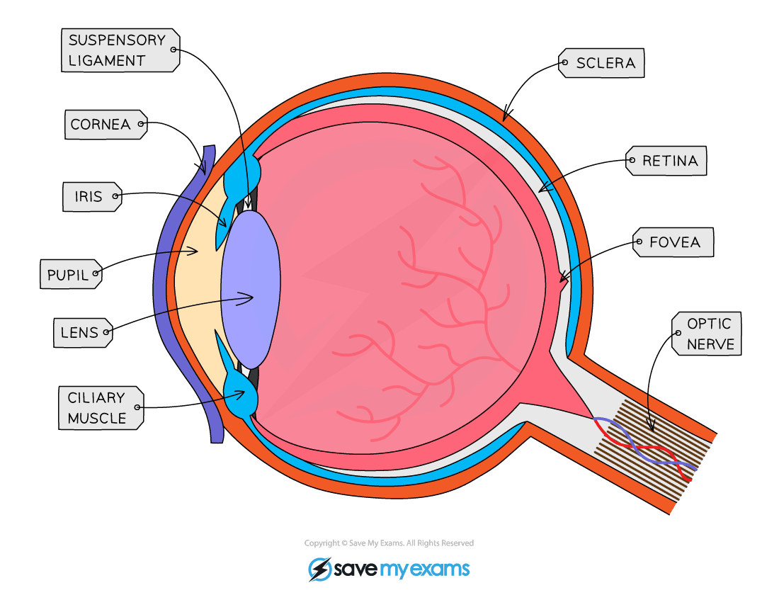
The Human Eye: Structure
- The eye is a highly specialised sense organ containing receptor cells that allow us to detect the stimulus of light
- The retina of the eye contains two types of receptor cells:
- Receptor cells that are sensitive to light, known as rods, and receptor cells that can detect colour, known as cones
The eye
Functions of the Parts of the Eye Table
- Other structures of the eye include:
- The ciliary muscle – a ring of muscle that contracts and relaxes to change the shape of the lens
- The suspensory ligaments – ligaments that connect the ciliary muscle to the lens
- The sclera – the strong outer wall of the eyeball that helps to keep the eye in shape and provides a place of attachment for the muscles that move the eye
- The fovea – a region of the retina with the highest density of cones (colour detecting cells) where the eye sees particularly good detail
- The aqueous humour – the watery liquid between the cornea and the lens
- The vitreous humour – the jelly-like liquid filling the eyeball
- The choroid – a pigmented layer of tissue lining the inside of the sclera that prevents the reflection of light rays inside the eyeball
- The blind spot – the point at which the optic nerve leaves the eye, where there are no receptor cells
The Human Eye: Function
The function of the eye in focusing on near and distant objects
- The way the lens brings about fine focusing is called accommodation
- The lens is elastic and its shape can be changed when the suspensory ligaments attached to it become tight or loose
- The changes are brought about by the contraction or relaxation of the ciliary muscles
- When an object is close up:
- The ciliary muscles contract (the ring of muscle decreases in diameter)
- This causes the suspensory ligaments to loosen
- This stops the suspensory ligaments from pulling on the lens, which allows the lens to become fatter
- Light is refracted more
Diagram showing the eye when an object is close up
- When an object is far away:
- The ciliary muscles relax (the ring of muscle increases in diameter)
- This causes the suspensory ligaments to tighten
- The suspensory ligaments pull on the lens, causing it to become thinner
- Light is refracted less
Diagram showing the eye when an object is far away
Focussing on Distant and Near Objects Table

The function of the eye in responding to changes in light intensity
- The pupil reflex is a reflex action carried out to protect the retina from damage
- In dim light, the pupil dilates (widens) in order to allow as much light into the eye as possible to improve vision
- In bright light, the pupil constricts (narrows) in order to prevent too much light from entering the eye and damaging the retina
The pupil reflex
The pupil reflex in dim light
The pupil reflex in bright light
Responding to Changes in Light Intensity Table
Exam Tip
The focusing of the eye on distant and near objects is complex and it can be hard to remember what is happening. This is something you can work out in an exam if you have forgotten – staring at your hand right in front of your eye will make your eyes feel tight and tired after a few seconds. This is because the ciliary muscles are contracted. Staring at an object far away feels relaxing and comfortable because the ciliary muscles are relaxed.


